


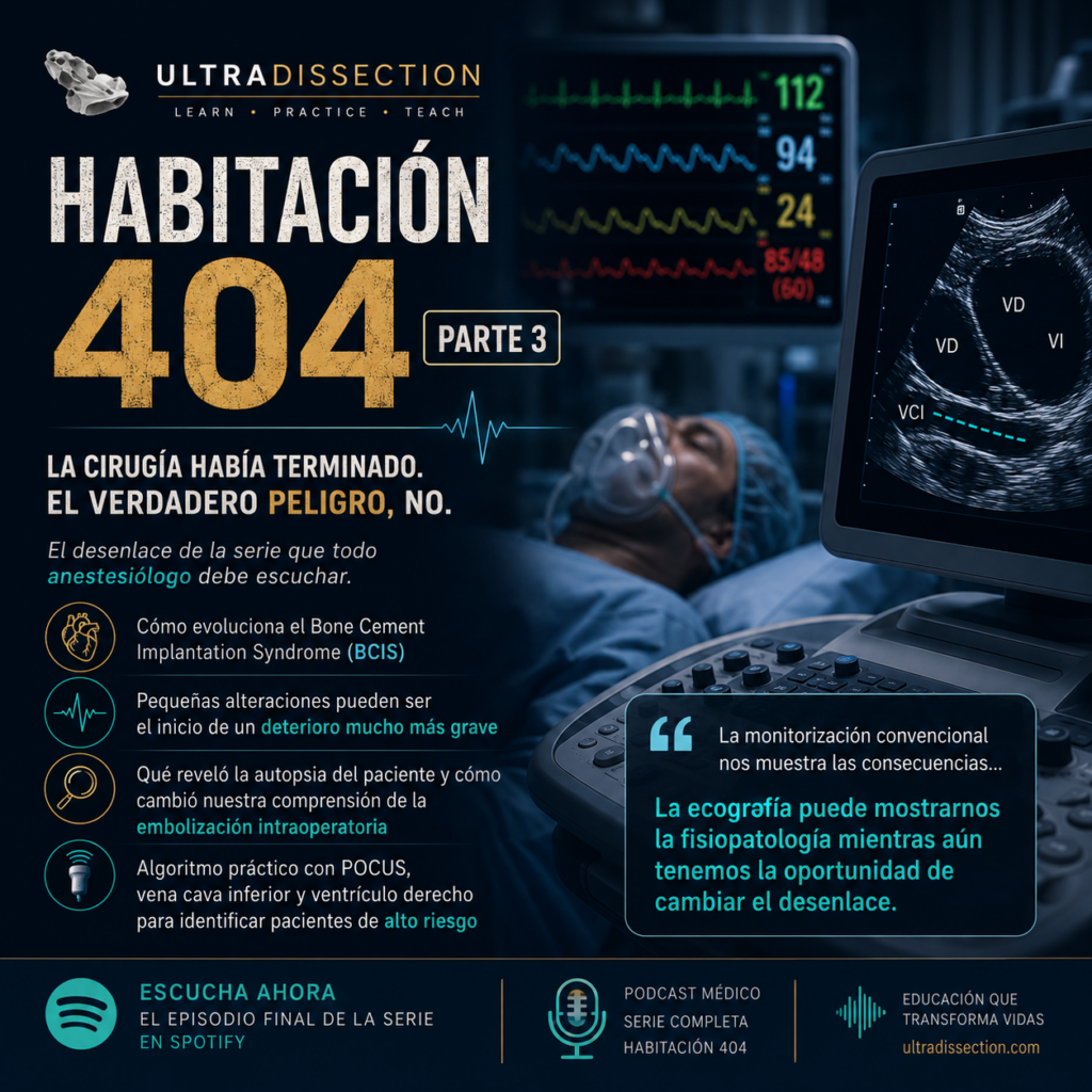
Cuando termina la cirugía… ¿realmente termina el problema?
La prótesis quedó correctamente implantada.
La herida fue cerrada.
El paciente estaba despierto, conversaba, respondía preguntas y la cirugía parecía haber concluido sin incidencias mayores.
En ese momento todo el equipo respiró tranquilo.
Sin embargo, la fisiopatología no entiende de tiempos quirúrgicos.
Aunque la intervención haya terminado, la embolización puede continuar, el ventrículo derecho puede comenzar a fatigarse y la circulación pulmonar puede deteriorarse de forma progresiva durante las siguientes horas.
Las pequeñas señales que solemos ignorar
Muchas complicaciones graves comienzan con cambios aparentemente insignificantes.
BCIS: el síndrome del cemento óseo
El Bone Cement Implantation Syndrome (BCIS) continúa siendo una entidad infradiagnosticada en muchos quirófanos.
Su presentación clínica puede ser muy variable.
Algunos pacientes desarrollan un colapso cardiovascular inmediato.
Otros presentan únicamente manifestaciones discretas que progresan de forma silenciosa durante las horas posteriores.
El paciente de la habitación 404
Cuatro horas después de la cirugía comenzaron a aparecer pequeños cambios.
- Desaturación leve.
- Taquicardia discreta.
- Hipotensión moderada.
- Necesidad de oxígeno suplementario.
Ninguno de estos datos parecía especialmente alarmante por sí solo.
Sin embargo, cuatro horas más tarde el paciente presentó una parada cardiorrespiratoria.
La autopsia
El diagnóstico definitivo fue contundente.
El nuevo algoritmo de actuación
Cuando exista embolización visible o aparezcan pequeños cambios clínicos durante una cirugía de cadera, no debemos normalizarlos.
Debemos iniciar una reevaluación sistemática.
Repetir la evaluación ecográfica.
Explorar el ventrículo derecho.
Valorar nuevamente la vena cava inferior.
Solicitar gasometría arterial.
Determinar troponinas y NT-proBNP.
Informar precozmente al equipo de Cuidados Intensivos.
Intensificar la monitorización.
Considerar una angio-TC pulmonar cuando esté indicada.
La verdadera enseñanza de este caso
Durante muchos años hemos monitorizado constantes vitales.
Hoy tenemos la oportunidad de monitorizar también la fisiopatología.
La ecografía nos permite observar procesos que comienzan mucho antes de que aparezca la hipotensión o la desaturación.
Ese cambio de paradigma puede modificar por completo la forma en que vigilamos a nuestros pacientes durante la cirugía ortopédica mayor.
Conclusión
La cirugía de cadera continúa siendo uno de los procedimientos con mayor potencial para producir fenómenos embólicos intraoperatorios.
Incorporar la evaluación ecográfica de la vena cava inferior y del ventrículo derecho durante los momentos críticos del procedimiento puede ayudarnos a identificar pacientes que requieren una vigilancia mucho más estrecha.
No se trata únicamente de reaccionar cuando el paciente colapsa.
Se trata de reconocer la fisiopatología mientras todavía tenemos la oportunidad de intervenir.
Comienzan a deteriorarse en el quirófano. La diferencia es que ahora contamos con herramientas que pueden ayudarnos a verlo antes.
Escucha la serie completa- Habitación 404.
Descubre cómo la ecografía perioperatoria está cambiando la manera en que entendemos la embolización intraoperatoria, el síndrome del cemento óseo y la monitorización fisiológica durante la cirugía de cadera.
▶ Escuchar en SpotifyWhen the Surgery Ends… Does the Risk End Too?
The prosthesis was successfully implanted.
The wound was closed.
The patient was awake, comfortable, answering questions, and everything appeared to have gone exactly as planned.
The operating room relaxed. The difficult case was finally over.
But physiology follows its own timeline.
Embolic material may continue traveling through the pulmonary circulation long after the final skin suture. Right ventricular strain may continue to increase. Pulmonary vascular resistance may continue rising. The pathological process does not stop simply because the operation has ended.
The Small Changes We Often Ignore
Many catastrophic complications begin with subtle physiological changes that are easily dismissed.
Bone Cement Implantation Syndrome (BCIS)
Bone Cement Implantation Syndrome remains one of the most underrecognized perioperative complications in orthopedic surgery.
Its presentation is highly variable.
Some patients deteriorate suddenly with immediate cardiovascular collapse.
Others develop only subtle abnormalities that progress silently over several hours before becoming life-threatening.
BCIS does not always present as a dramatic intraoperative collapse. Delayed clinical deterioration may be equally important.
The Patient in Room 404
Four hours after surgery, small changes began to appear.
- Mild oxygen desaturation.
- Subtle tachycardia.
- Moderate hypotension.
- Increasing oxygen requirements.
None of these findings appeared dramatic when considered individually.
Four hours later, the patient suffered cardiac arrest.
The Autopsy
The final diagnosis was unequivocal.
A New Clinical Algorithm
Significant embolization, unexplained hypoxemia, progressive tachycardia or hypotension should never be automatically attributed to spinal anesthesia or postoperative stress.
Instead, they should trigger a structured reassessment.
Repeat focused ultrasound examination.
Evaluate right ventricular function.
Reassess the inferior vena cava.
Obtain arterial blood gas analysis.
Measure cardiac biomarkers including Troponin and NT-proBNP.
Notify the intensive care team early.
Escalate monitoring and hemodynamic surveillance.
Consider CT Pulmonary Angiography when clinically appropriate.
The Most Important Lesson
For decades we have monitored vital signs.
Today we have the opportunity to monitor physiology itself.
Ultrasound allows us to observe pathological processes before they become hemodynamic disasters.
This represents a fundamental shift in perioperative medicine: moving from reacting to deterioration... to anticipating it.
Final Thoughts
Total hip arthroplasty remains one of the orthopedic procedures with the greatest potential for intraoperative embolization.
Incorporating focused assessment of the inferior vena cava and right ventricle during the critical phases of surgery may allow clinicians to recognize high-risk patients long before cardiovascular collapse occurs.
The goal is not to transform every anesthesiologist into an advanced echocardiographer.
The goal is much simpler: learn to recognize the physiological patterns that truly change clinical decision-making.
Perhaps they begin dying in the operating room— and we simply have not learned to recognize the earliest signs. Ultrasound may allow us to change that story.
Listen to the full series
Discover how perioperative ultrasound is transforming our understanding of intraoperative embolization, Bone Cement Implantation Syndrome (BCIS), and physiological monitoring during total hip arthroplasty.
▶ Listen on Spotify