


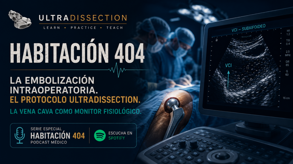
La cirugía de cadera es una cirugía embolígena
Aunque pocas veces se menciona durante la formación anestésica, cada artroplastia de cadera representa un escenario con potencial embolígeno importante.
Cada vez que el cirujano fresa el canal femoral, realiza un lavado medular, cementa o introduce el vástago protésico, la presión intramedular aumenta de forma considerable.
Cuando esa presión supera la capacidad del canal óseo, el contenido medular encuentra una vía de salida hacia la circulación venosa.
La verdadera pregunta es: ¿estamos siendo capaces de verla mientras ocurre?
¿Qué entra realmente en la circulación?
Con frecuencia hablamos únicamente de embolia grasa. Sin embargo, la realidad es mucho más compleja.
Lo que asciende hacia la circulación pulmonar puede contener una mezcla de diferentes materiales procedentes del canal medular.
En conjunto forman una auténtica tormenta embólica, capaz de alterar progresivamente la circulación pulmonar.
Los momentos donde debemos mirar
La mayoría de los anestesiólogos incrementa su atención cuando aparecen cambios en el monitor.
Sin embargo, la vigilancia debería aumentar antes, exactamente durante los momentos con mayor riesgo de embolización.
- Luxación de la cadera.
- Fresado femoral.
- Lavado del canal medular.
- Cementación.
- Inserción del vástago.
- Reducción final.
Si esperamos a que aparezca la hipotensión, probablemente ya hemos perdido la oportunidad de observar el inicio del proceso fisiopatológico.
La publicación que cambió nuestra forma de pensar
Un grupo canadiense demostró algo extraordinario.
Durante una artroplastia de cadera realizada bajo anestesia espinal utilizaron una sonda convexa para observar la vena cava inferior en tiempo real.
Primero aparecieron pequeñas partículas.
Después una lluvia embólica.
Finalmente una auténtica tormenta de material embólico.
Solo después aparecieron las alteraciones hemodinámicas.
La vena cava nos está hablando
No necesitamos un ecocardiograma transesofágico.
No necesitamos una unidad de hemodinámica.
Tampoco hace falta ser cardiólogo.
Necesitamos una sonda convexa, conocer la anatomía y saber interpretar patrones sencillos.
Protocolo Ultradissection
Monitorización ecográfica de la vena cava durante cirugía de cadera
Obtener una imagen basal antes de comenzar cualquier manipulación del canal femoral.
Utilizar un abordaje subxifoideo con sonda convexa de 2–5 MHz.
Identificar correctamente:
- Hígado.
- Vena cava inferior.
- Aurícula derecha.
Medir aproximadamente entre 1 y 2 cm antes de la aurícula derecha y registrar la imagen basal.
Repetir la exploración durante:
- Luxación.
- Fresado.
- Cementación.
- Inserción del vástago.
- Reducción.
¿Qué estamos buscando?
La imagen característica recuerda una tormenta de nieve moviéndose dentro de la circulación.
Pequeños ecos brillantes, móviles, ascendiendo por la vena cava hasta alcanzar la aurícula derecha.
El siguiente paso: mirar el ventrículo derecho
Cuando observamos una embolización importante, la siguiente estructura que debemos explorar es el corazón derecho.
No buscamos cifras sofisticadas.
Buscamos respuestas sencillas.
- ¿El ventrículo derecho está dilatado?
- ¿Es igual o mayor que el izquierdo?
- ¿El septo se encuentra aplanado?
- ¿La cavidad izquierda adquiere forma de "D"?
- ¿La vena cava permanece dilatada y sin colapsar?
Interpretar antes de reaccionar
El verdadero cambio de paradigma consiste en dejar de reaccionar únicamente cuando aparece el colapso.
Si la ecografía nos demuestra que la embolización ya está ocurriendo, disponemos de minutos valiosos para intensificar la vigilancia, preparar soporte avanzado y anticiparnos al deterioro.
Lo complementa mostrando la fisiopatología mucho antes de que aparezcan las consecuencias.
Escucha la serie especial, Habitación 404
Descubre cómo la ecografía perioperatoria está cambiando la manera en que entendemos la embolización intraoperatoria, el síndrome del cemento óseo y la monitorización fisiológica durante la cirugía de cadera.
▶ Escuchar en SpotifyHip Arthroplasty Is an Emboligenic Procedure
Although it is rarely emphasized during anesthesia training, every total hip replacement carries a significant risk of intraoperative embolization.
Every time the femoral canal is reamed, irrigated, cemented, or the prosthetic stem is inserted, intramedullary pressure rises dramatically.
Once that pressure exceeds the capacity of the medullary canal, its contents are forced into the venous circulation.
The real question is: Are we able to recognize it while it is happening?
What Actually Enters the Circulation?
We often refer simply to fat embolism. In reality, the embolic load generated during hip arthroplasty is considerably more complex.
Multiple types of material may be released into the venous system during femoral canal manipulation.
Together, these elements create what can only be described as an embolic storm, capable of progressively increasing pulmonary vascular resistance and placing significant strain on the right ventricle.
The Moments That Matter Most
Most anesthesiologists become highly attentive only after physiological changes appear on the monitor.
However, surveillance should actually intensify during the surgical stages that generate the highest embolic load.
- Hip dislocation.
- Femoral canal reaming.
- Medullary canal irrigation.
- Bone cement application.
- Stem insertion.
- Final reduction.
If we wait for hypotension or oxygen desaturation, we may already have missed the earliest phase of the underlying pathophysiology.
The Study That Changed Our Perspective
A Canadian research group demonstrated something remarkable.
During total hip arthroplasty performed under spinal anesthesia, they continuously visualized the inferior vena cava using a convex ultrasound probe.
Initially, only a few isolated echogenic particles appeared.
Moments later, the number increased into what resembled a shower of embolic material.
Finally, an unmistakable embolic storm filled the vena cava before any significant hemodynamic deterioration became evident.
The Inferior Vena Cava Is Speaking to Us
We do not need transesophageal echocardiography.
We do not need an interventional cardiology suite.
We do not even need to become expert echocardiographers.
We simply need a convex probe, a basic understanding of anatomy, and the ability to recognize clinically relevant patterns.
The UltraDissection Protocol
Inferior Vena Cava Monitoring During Hip Arthroplasty
Obtain a baseline ultrasound image before any manipulation of the femoral canal.
Use a 2–5 MHz convex probe through a subxiphoid approach.
Clearly identify:
- The liver.
- The inferior vena cava.
- The right atrium.
Measure the vena cava approximately 1–2 cm before its entrance into the right atrium and record the baseline image.
Repeat the examination during:
- Hip dislocation.
- Femoral reaming.
- Cementation.
- Stem insertion.
- Final reduction.
What Are We Looking For?
The characteristic ultrasound appearance resembles a snowstorm moving through the venous circulation.
Bright mobile echoes ascend through the inferior vena cava toward the right atrium like countless tiny particles suspended in flowing blood.
The Next Step: Assess the Right Ventricle
Once significant embolization has been identified, attention should immediately shift toward evaluating right ventricular function.
Sophisticated quantitative measurements are not required.
Simple clinical questions often provide the information we need.
- Is the right ventricle dilated?
- Is it equal to or larger than the left ventricle?
- Has the interventricular septum flattened?
- Does the left ventricle appear D-shaped?
- Is the inferior vena cava enlarged and poorly collapsible?
Recognize Before You React
The true paradigm shift is learning to identify the pathophysiological process before cardiovascular collapse occurs.
If ultrasound demonstrates embolization while the patient remains apparently stable, valuable minutes are gained to intensify monitoring, prepare advanced support, and anticipate deterioration rather than simply reacting to it.
It complements it by revealing the underlying physiology long before traditional monitors display its consequences.
Listen to the three part series, Room 404
Discover how perioperative ultrasound is transforming our understanding of intraoperative embolization, Bone Cement Implantation Syndrome (BCIS), and physiological monitoring during total hip arthroplasty.
▶ Listen on Spotify